Microscopy is the workhorse of the physical and life sciences, producing crisp images of everything from atoms to cells well beyond the capabilities of the human eye. However, the analysis of these images is frequently little better than automated manual marking. Here, we revolutionize the analysis of microscopy images, extracting all the information theoretically contained in a complex microscope image. Using a generic, methodological approach, we extract the information by fitting experimental images with a detailed optical model of the microscope, a method we call Parameter Extraction from Reconstructing Images (PERI). Our focus here is on images of colloidal images taken with a confocal microscope, but the ideas here are broadly applicable to any form of microscopy.
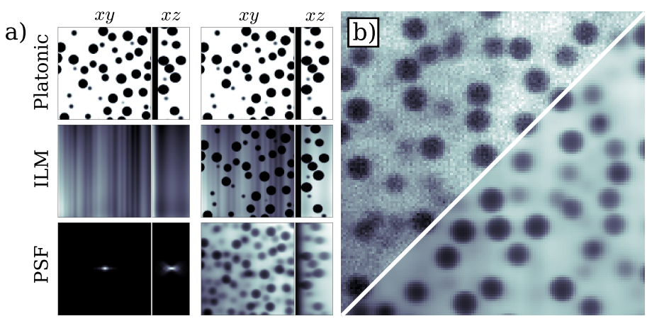
Below, you can observe the process by which our generative model creates colloidal data. Because we are primarily focused on tracking spherical particals, our model images begin with a Platonic image of dye distributed around perfects spheres (a, top row). Our model goes on to incorporate pixelation, as well as detailed estimates of the spatially-varying illumination created by the microscope light source (a, middle row) and the point spread function (a, bottom row). Finally, we add a realistic noise level to complete the picture. Panel b) shows real data in the upper left corner, and our generated data (without added noise), and by eye it is already clear that these iimages match well. When we add noise to the bottom right image, the two become virtually indistinguishable.
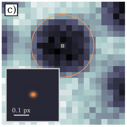
Using our generative model, we are able to locate the size and position of micron-sized particles with nanometer precision. This level of measurement is very near the information-theoretic bound on how accurately these quantities could possibly be measured. Below is a zoomed-in image of a single spherical particle; the inset shows the grey box at the center of the particle, and withing that the estimate of the particle's center from PERI. The estimates illustrated here are on the order of a few percent of a pixel, which translates to a few nanometers for these images!
With this unprecedented level of measurement precision, we open the door to a whole new suite of analyses for colloidal systems that rely on extreme measurement precision. Moreover, the methods used in PERI are broadly applicable, and can be extened to a wide range of systems. Stay tuned for these future applications!
Pre print: https://arxiv.org/abs/1702.07336
PERI code and tutorial: http://www.lassp.cornell.edu/sethna/peri/index.html
Github: https://peri-source.github.io/peri-docs/